 At Unilaser Médica, we treat the different presentations of visible veins on the skin using laser, IPL, and radiofrequency. Visible vessels have very diverse presentations.
Factors to Consider in Treatment
1. Vessel Density
Facial telangiectasias vary in density. In cases of high density, such as photodamage or rosacea, multiple sessions are required to treat the abundance of very varied thickness types.

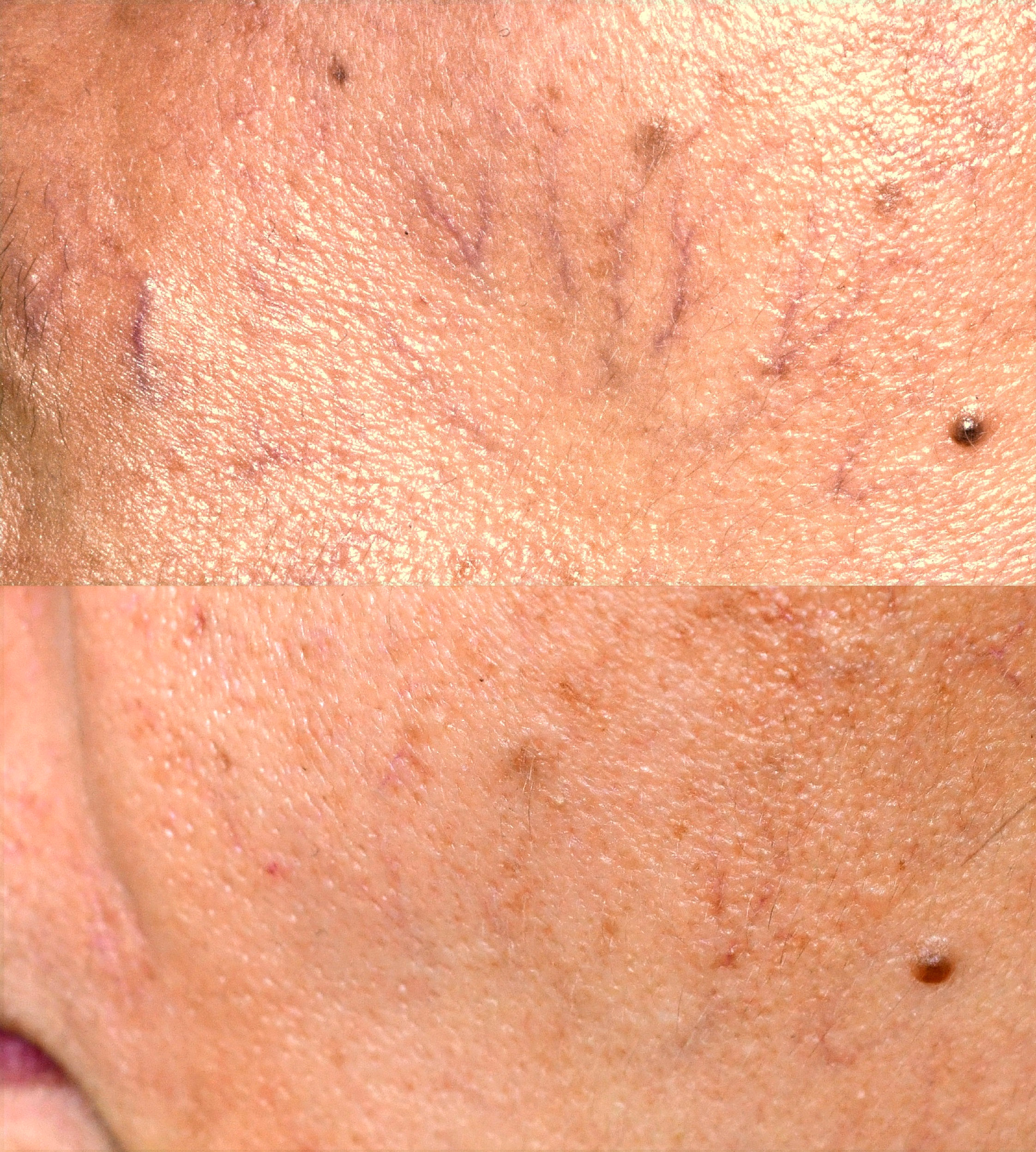
Result with the Fraxel 1550 laser in the treatment of poikiloderma. Appearance at 10x magnification of skin with photodamage (vascular dilations and pigment structures are observed).
2. Vessel Thickness
Vessel thickness may require different types of lasers or power and pulse length parameters.

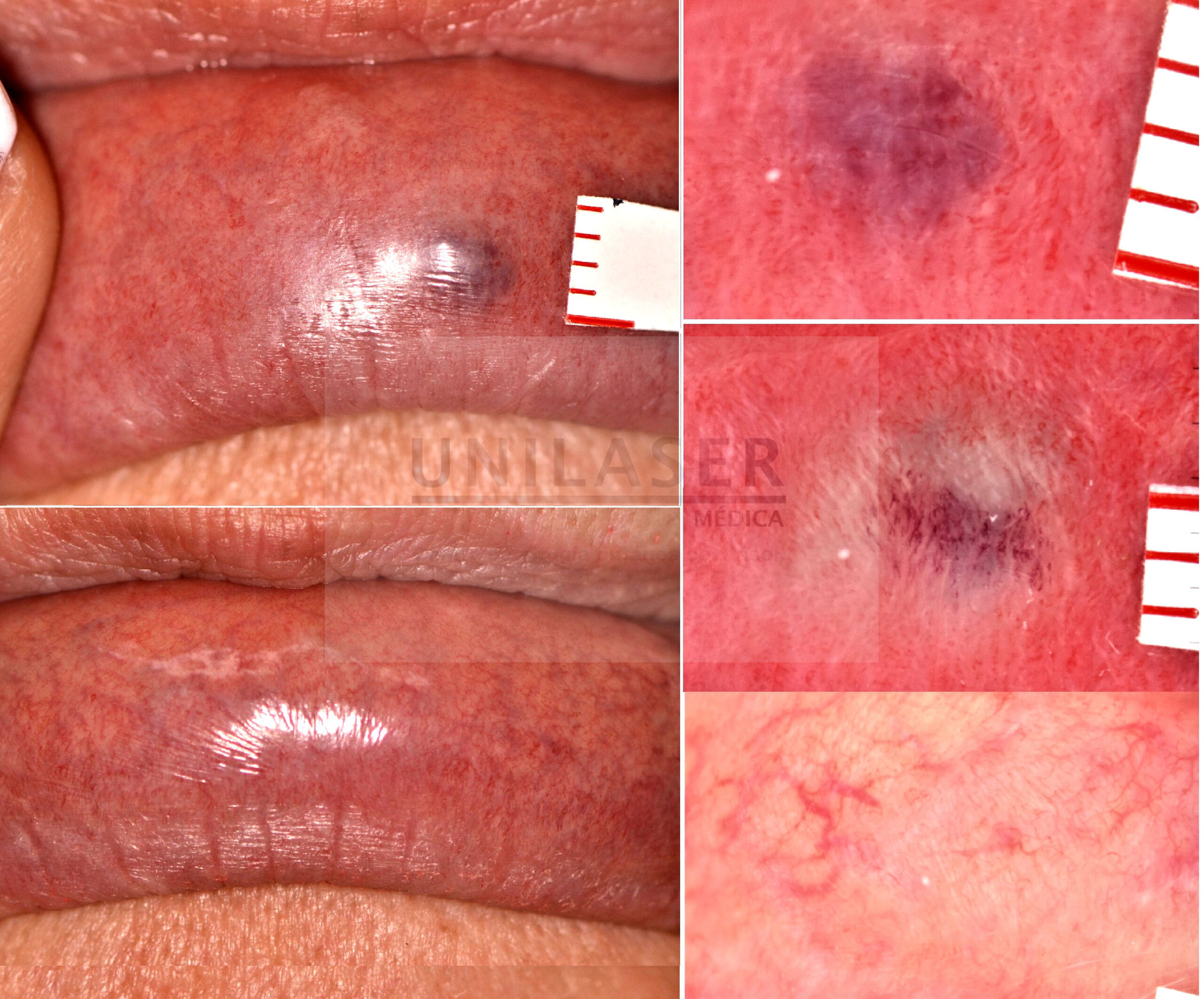
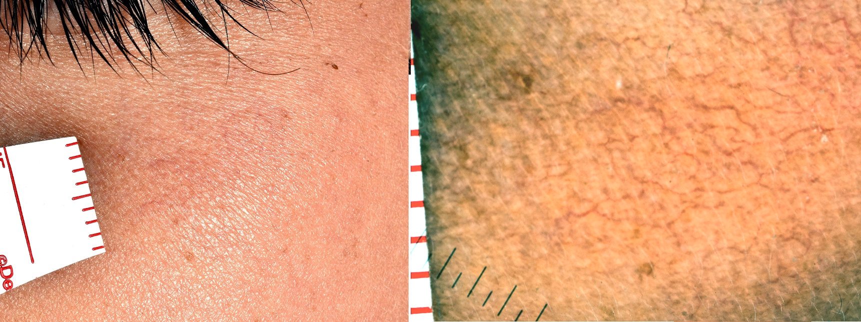
Superficial violet vessels of the upper lip.
3. Vessel Depth
Depth affects laser choice. Lasers like Nd-YAG are effective for deeper vessels. Sometimes it may also be necessary to choose direct access with radiofrequency.

Violet vessels at usual depth.
Types of Laser and Their Application

Results of the Nd YAG Primelase laser on facial telangiectasias
Primelase Nd-YAG® Laser: Ideal for treating violet and red colored vessels. It penetrates deeply to coagulate the affected vessels. (See gallery: Top photo violet vessels of medium depth / Bottom photo result at 4 months).
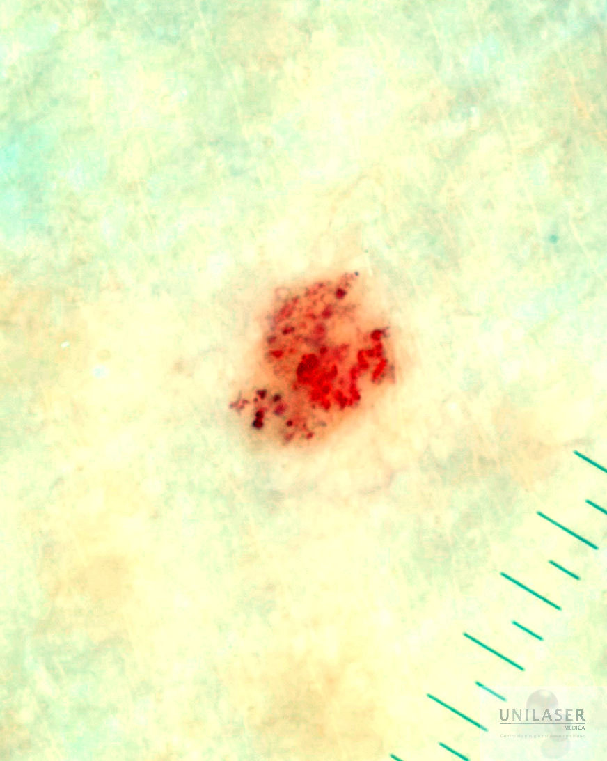
- Milesman® 445 Laser: Used for finer and more superficial vessels. It offers high selectivity. (See gallery: Partial result on multiple chest lesions of a 90-year-old woman).
- Viridex®: Ultra-modulated radiofrequency equipment. It works by direct contact with the vessel.


Important Considerations
Multiple Sessions
Most cases require several sessions to achieve complete results. (Example: 4-session treatment over a 6-month period).
 – copia")
Post-Treatment Follow-up
Regular follow-up is essential to evaluate the efficacy of the treatment and make adjustments if necessary.
Risks and Safety
It is important to discuss potential risks and post-treatment care. The collapse of the laser-treated vessel leaves the space it occupied in the skin intact.

Vessel Density: A Key Factor
The presence of numerous telangiectasias on the face implies a detailed approach to treat a variety of vessels, from the finest to the thickest, located at different depths.
Hemangiomas and Major Lesions
Smaller and more segmented dilations, such as cherry angiomas, are easier to treat due to their greater circumscription and small size. Slightly larger lesions with larger diameter vessels respond better to Polaris, a laser that adds radiofrequency to the shot.
Sequence after shot with Polaris RF equipment of venous lake

Immediate effect of intense pulsed light on dilated vessels of the nasal wing.
 Cherry angiomas (red dots or balls) are small, superficial, and very well circumscribed. Coagulation is immediate with little pain and better recovery using lasers.
Treatment with the Milenium 450 laser is especially beneficial when we have a high quantity of very small cherry angiomas on the body. More specific lesions can be treated quickly without the use of anesthesia.
When treating the vessel of origin of the spider vein, it may appear as ineffective coagulation to the naked eye, but it is demonstrated by dermoscopy. Thrombosis of the feeding vessel allows the lesion to disappear weeks later without additional treatment. (Ex: 45 days later, the thrombosis of the main vessel resolved, and so did the spider nevus).
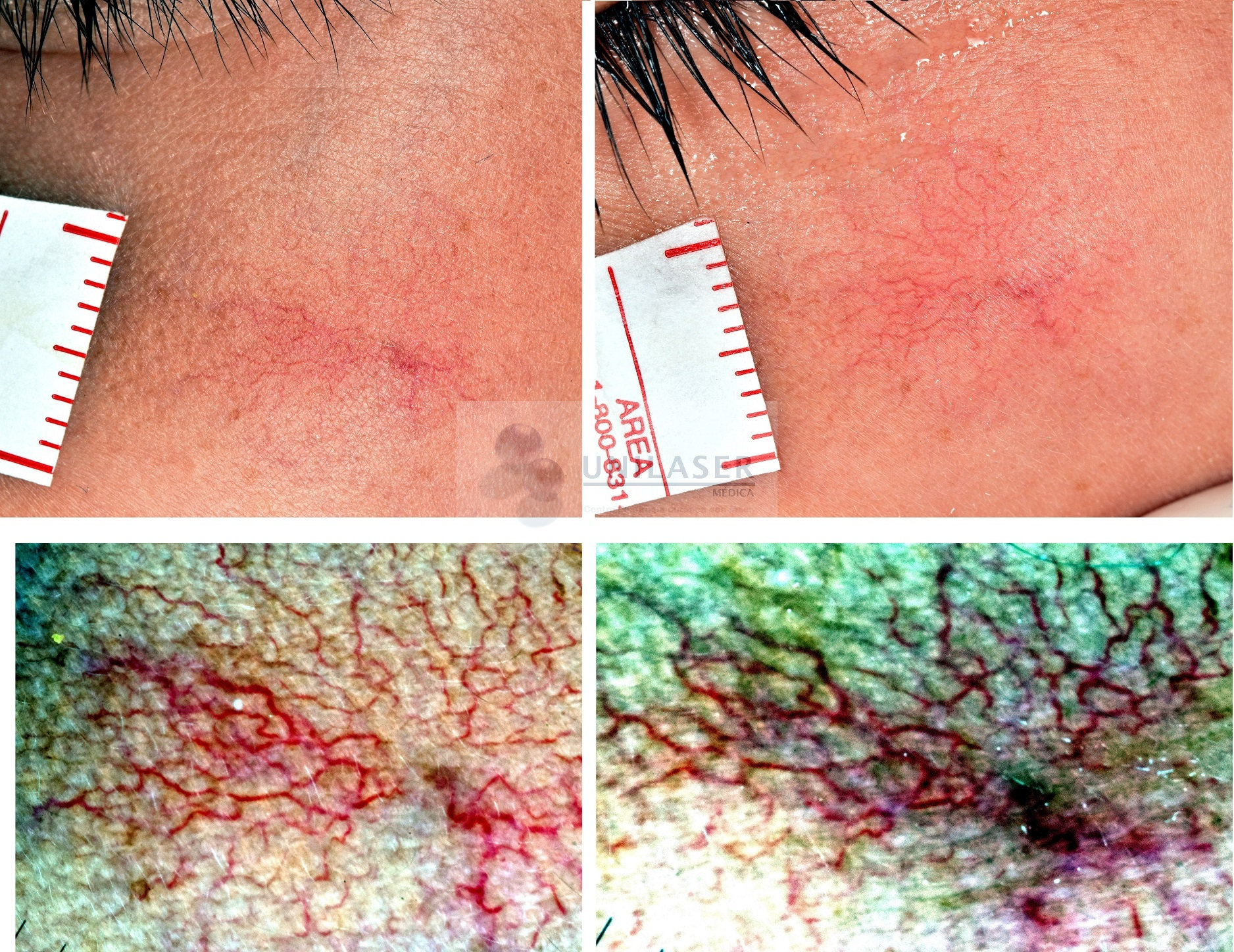
The main vessel seems even more noticeable on day 45 after the first session but with apparent thrombosis. He did not fall into the temptation of looking for the visible collapse since it could have caused a destruction of the skin and the lesion a month and a half later, resolved spontaneously.
Result 45 days later with disappearance of the nutrient vessel and a good part of the telangiectasias