
Laser Removal of Skin Cysts
The removal of skin cysts using a laser, under microscopic assistance, prevents skin depressions.
What are skin cysts?
They can be small, numerous bumps that appear on the skin of the face, or larger ones that usually appear on the rest of the body, especially on the back. They are formed by the “entrapment” of keratin, which is the protein that forms the skin and that we shed every day. For this reason, they are named “epidermoid cysts.”
How are skin cysts removed with laser?
Some very small ones can disappear spontaneously or through very discreet manipulation with needles. When they are deeper and larger, they displace neighboring tissues and can adhere to the skin segment where they originated, occasionally showing a small opening. Surgical resection is the norm. Some practitioners prefer to inject them with steroids, but complications such as atrophy and skin depigmentation, increased blood vessels, and persistence of the cyst can occur.
Why is laser useful for removing skin cysts?
In smaller cysts, they can be unroofed and extracted; in larger ones, the laser facilitates dissection and extraction by better respecting the edges to close the incision. In larger ones, complete excision of the capsule that envelops the cyst is necessary. Microscopic dissection and bleeding control allow for a cleaner and clearer resection.
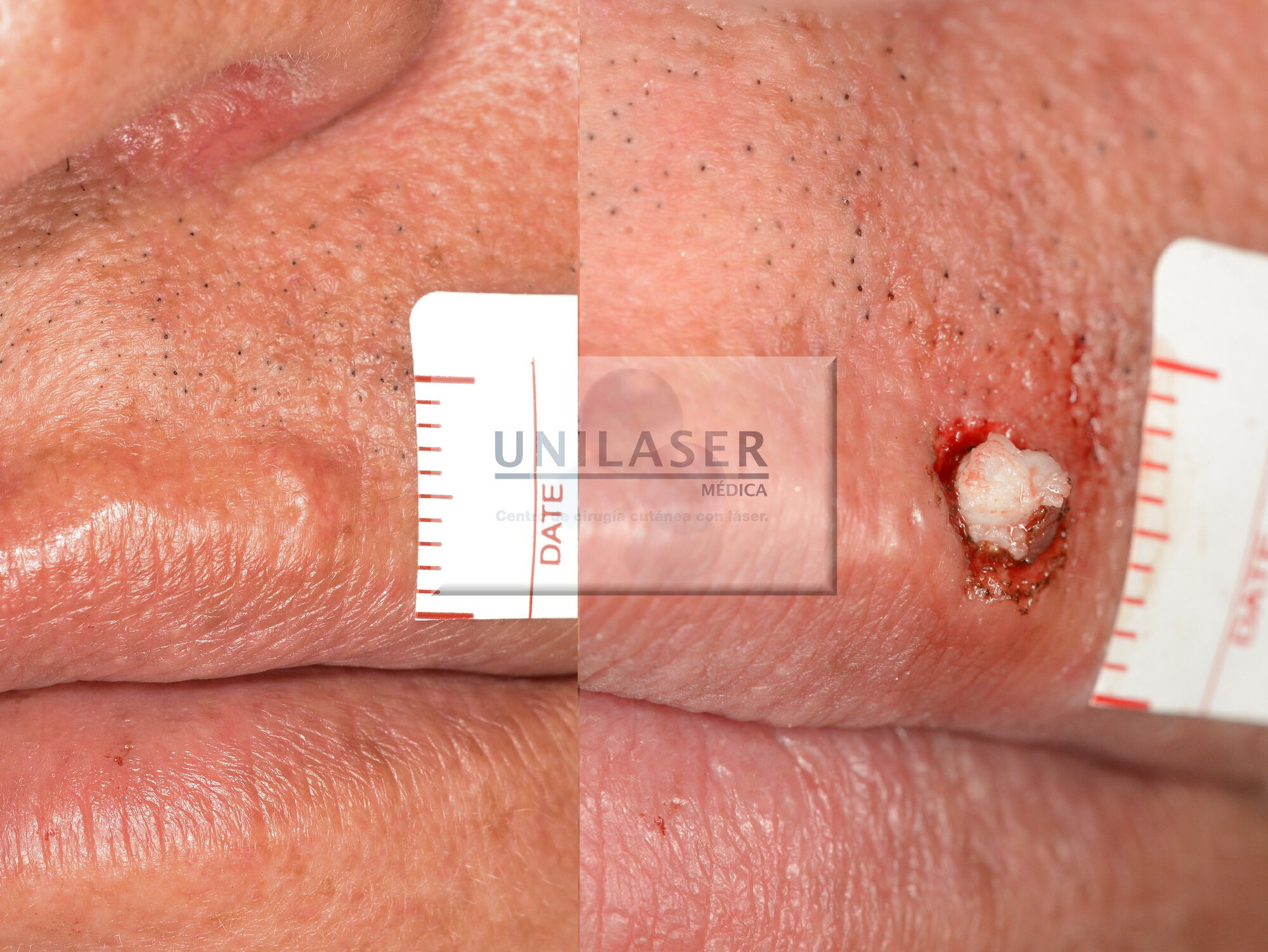
Quiste epidermoide del labio
CO2 laser dissection of epidermoid cyst
CO2 laser dissection of epidermoid cyst.
What to do when the cyst is inflamed?
In these cases, the purulent material must be drained, and a gauze “wick” must be placed and changed daily until closure is ensured.
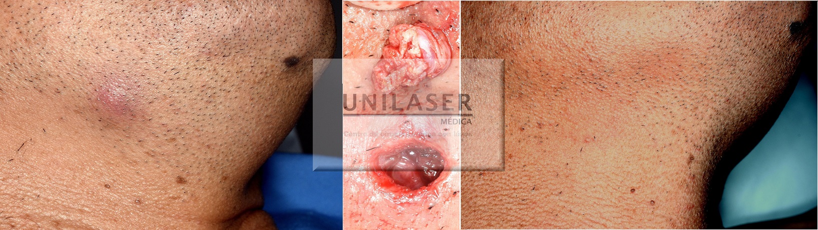
quiste inflamado drenaje y extracción
Closure by secondary intention can sometimes leave a better scar despite having to perform a greater number of healings.
Closure by secondary intention can sometimes leave a better scar despite having to perform a greater number of healings.
Why do scalp cysts have to be operated on?
They grow and sometimes are more numerous. They tend to be recurrent even after surgery.
When do vellus cysts appear?
They generally appear on the trunk of adolescents and young adults. They are characterized by having vellus hair shafts inside the cysts and are called eruptive vellus hair cysts. A solitary vellus-type cyst with terminal hair can occur, in most cases only on the head and neck.
Are there congenital cysts?
They are especially located around the orbits, and their capsule contains skin appendages. The hair shafts are of the lanugo type. They are called dermoid cysts.
Cysts on the eyelids?
Epidermoid types form easily, conditioned by frequent scratching or rubbing. It is not uncommon to see hidrocystomas, which are transparent and ideal for intervention with laser given their thin capsule. Laser-assisted extraction of epidermoid cysts with laser.
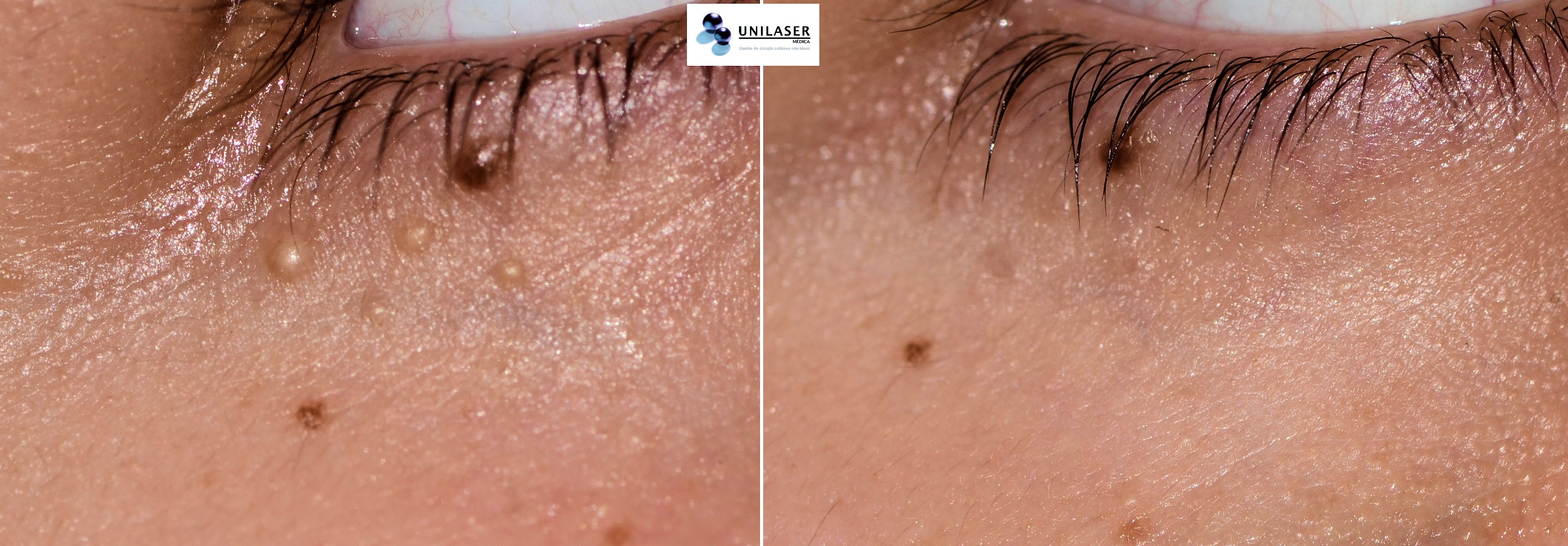
Destechamiento y vaporización de las cápsulas asitida con láser
Laser-assisted deroofing and vaporization of capsules
Laser assisted emptying and vaporization of capsules.
Milia
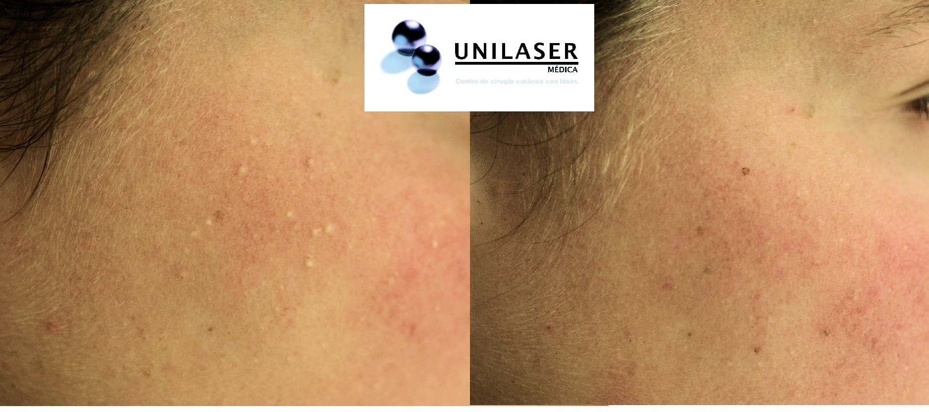
Destechamiento, vaporización de microcapsulas asistida con láser y microscopio
Laser and microscope assisted deroofing, vaporization of microcapsules
Extraction of milium. No scars observed after 8 days.
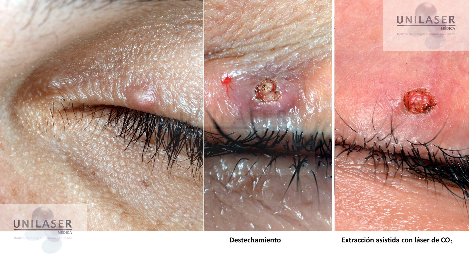
Extracción quiste epidermoide palpebral
Visualization of two keratin clumps, laser-assisted extraction allows leaving a clean bed for closure
Manual puncture of the capsule vs manual puncture + vaporization of the base using Er:YAG laser. Note the difference.
